A Novel Training Method for Endovascular Clot Retrieval Using a Portable Vascular Model and Red Film
Article information

Abstract
Hands-on training is a crucial part of education in neuroendovascular treatment to ensure safe and rapid acquisition of techniques. However, there is a significant gap between training and actual clinical practice. This study will introduce innovations for more practical thrombus retrieval training that was developed in this process. A Smart Vascular Model 3 in 1 was used. A pink pseudothrombus was inserted into the M1 (horizontal segment of the middle cerebral artery) section of the model. Then, a “red underlay” purchased at a stationery store was placed to cover the proximal part of M1 and beyond so that the pseudothrombus was not visible. The thrombus was retrieved during training by looking for the location of the thrombus based on the behavior and resistance of the tip of the guidewire and deployment of the stent retriever. The participants were required to have detailed observation skills and precise manipulation skills using a red film to prevent the direct visualization of the pseudothrombus. The implementation of this innovation to the previous hands-on training made the training more practical and effective. If the exact thrombus location can be determined by the behavior of the wire tip, the device’s capabilities can be maximized, and rapid retrieval can be expected. It could also reduce complications, as unnecessary peripheral guidance of the device could be avoided.
INTRODUCTION
Hands-on training in endovascular therapy is necessary because it introduces beginners to new medical devices and allows them to explore the limitations of techniques and equipment. However, during conventional hands-on training, all that can be learned in the limited time available is how to set up and use the equipment. In particular, training in endovascular clot-retrieval procedures has a significant discrepancy with clinical procedures. Without radioscopy, the pseudothrombus in a vascular model can be confirmed under direct visualization, making the training too easy.
Previously, portable vascular models used for training in multiple procedures including clot retrieval were developed [1]. This study presents a novel hands-on training method that was found to be more practical, educational, and simpler than conventional methods.
MATERIALS AND METHODS
The resinous A4 sized Smart Vascular Model 3 in 1 (Nihon Light Service Inc.) was used (Fig. 1A). This vascular model has 2 ports on the back that can be used to inject pseudoclots. The pseudoclots were made by mixing commercially available borax, laundry glue (containing polyvinyl alcohol), red water-based paint, and water. However, in conventional hands-on training using these devices, the thrombus was revealed under direct vision.
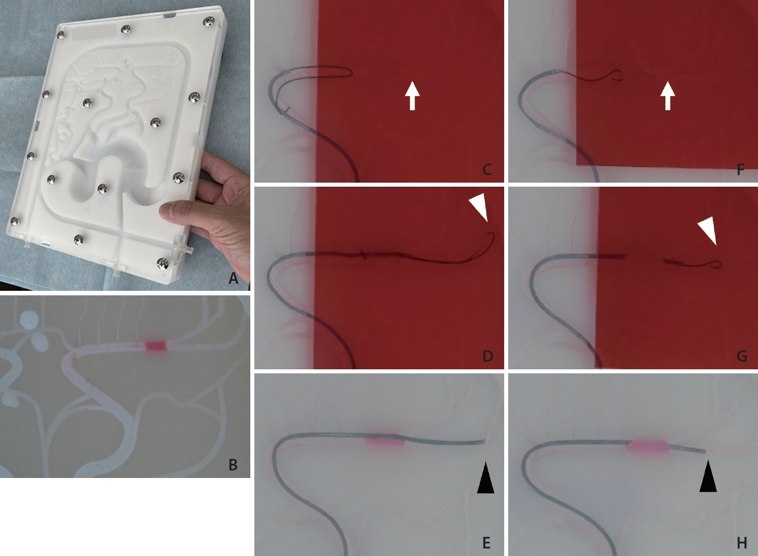
(A) Smart Vascular Model 3 in 1 (Nihon Light Service Inc.). (B) A pink pseudoclot was placed in the vessel. (C–E) Vascular model during clot retrieval using a large J-shaped guidewire. (C) When the red film was covered, the pseudothrombus became completely invisible (white arrow). A large J-shaped guidewire was used to pass through the blockage. (D) The white arrowhead indicates the tip of the curved guidewire. (E) The microcatheter was navigated into the far distal vessel (arrowhead). (F–H) Vascular model during clot retrieval using a small J-shaped guidewire. (F) When the guidewire contacted the invisible pseudothrombus (white arrow), it deflected, and the origin of the thrombus could be ascertained. (G) Once the guidewire passed through the thrombus and reached the true lumen of the vessel, the wire tip was free (white arrowhead), and the distal end of the thrombus could be recognized. (H) The microcatheter could be guided into the appropriate position (arrowhead).
Therefore, we used a red underlay bought at a stationery store. When the red film covered the area distal to the M1 (horizontal segment of the middle cerebral artery) origin, the location of the pseudothrombus was completely obscured (Fig. 1B, C). A Phenom 21 microcatheter (Medtronic) and a CHIKAI 14 guidewire (Asahi Intecc) were used to penetrate the clot. A Solitaire 4×40 (Medtronic) was used for clot retrieval. The inside of the vascular model was not filled with water. The inside of the catheter was rinsed with water once.
RESULTS
Once the red film covered the pseudoclot, the location of the clot was completely obscured. Rather, the behavior of the wire, position of the microcatheter, and deployed stent retriever were observed.
When the wire tip entered in a large J-shape with an approximately 20-mm bend (Fig. 1C), it was impossible to recognize the proximal end of the thrombus, and even when the distal lumen of the thrombus was reached, it was impossible to determine the distance to the actual thrombus (Fig. 1D). As a result, the microcatheter was guided more distally than was necessary (Fig. 1E).
In comparison, when the tip of the wire was made into a small J-shape of approximately 2.5 mm and advanced into the thrombus (Fig. 1F), the deflection and resistance of the wire made the proximal end of the thrombus palpable (Fig. 1G). Furthermore, once the wire tip reached the true lumen of the vascular model from the distal end of the thrombus, the wire was suddenly free, and the position of the distal end of the thrombus could be determined. Thus, the microcatheter could be guided to the appropriate position (Fig. 1H).
DISCUSSION
Hands-on training for endovascular procedures is essential to master the correct and safe use of devices. Specifically, simulation using a vascular model is important because a rapid procedure is required to achieve good results in mechanical clot retrieval. In addition to the traditional face-to-face training between instructor and participant, the effectiveness of online hands-on training has been reported during the recent coronavirus disease 2019 pandemic [1]. However, several problems exist with hands-on training in clot retrieval procedures. The most urgent problem is that hands-on training is very different from actual clinical procedures. This is because the exact location of the thrombus is not known in actual clinical practice. It would be nice if training could be conducted under fluoroscopic guidance; however, finding a place and time for several participants is difficult, and radiation exposure for both the instructor and trainees is also a problem.
We devised a method to make the red clots invisible under direct vision by introducing a red underlay used by students studying for a test. The clot was not visible; however, the behaviour of the wire and catheter could be seen. In actual clinical practice, even with sandwich contrast injection from a guiding catheter and microcatheter, it is difficult to delineate the exact location of a thrombus. The thrombus tends to be overestimated. Therefore, covering the distal side from the M1 proximal point with a red film could create conditions similar to those in actual clinical practice.
Some authors have reported the effectiveness of using small J-shaped microwires for clot retrieval [2,3]. As presented in this study, when the tip of the guidewire contacted the proximal end of the thrombus in a large J-shape, the shape of the wire did not change at all, nor was it transmitted as resistance to the surgeon. The shape of the wire also did not change when the tip of the wire crossed the distal end of the thrombus and reached the true lumen of the vessel. This tended to guide the wire and microcatheter much further distally than the thrombus. Reaching unnecessarily far distally increases the risk of vascular injury by the wire and stent retriever.
On the contrary, with a small J-shaped wire, the exact location of the thrombus could be determined. Once the exact location of the thrombus is known, the sweet spot of the stent retriever can capture the thrombus, and the aspiration catheter can provide reliable contact aspiration. This is expected to improve the retrieval success rate. In conventional hands-on training, the technique is performed while somehow looking at the clot location; however, by applying the red film, the training becomes more like clinical practice and requires a high level of concentration and skill.
This study has some limitations. First, given that this method does not allow imaging with contrast media, a discrepancy was observed in the information obtained in actual clinical practice. Second, regarding the pink pseudothrombus used in this study, if the device happens to pass through the front side of the thrombus, an image similar to that obtained under fluoroscopy can be obtained, as shown in Fig. 1D. On the other hand, if the device passes through the back side of the thrombus, as shown in Fig. 1G, the thrombus shadow is defective and the location of the thrombus is known. To solve this problem, we created a transparent pink pseudothrombus. Third, the Smart Vascular Model 3 in 1 used in this study is a 2-dimensional (2D) vascular model; thus, training was performed on an anteroposterior image only. In a 3D vessel, the findings of wire behavior should be different. In the future, we plan to develop 3D models and models with more ports into which thrombi can be inserted. Finally, to demonstrate the purpose of this study, a comparison with conventional training methods should be made.
CONCLUSION
The addition of this innovation to our existing hands-on training makes it more practical and effective. If the exact location of the thrombus can be determined by the behaviour of the wire tip, the device’s capabilities can be maximized, and rapid retrieval can be expected. It may also contribute to a reduction in complications, as unnecessary peripheral guidance can be avoided. Thus, improving participants’ techniques can contribute to the development of safer and faster neuroendovascular treatment.
Notes
Fund
None.
Ethics Statement
This study waived approval of the Institutional Review Board (IRB). The consent for publication is not required as the submission does not include any images or information that may identify the person.
Conflicts of Interest
The authors have no conflicts to disclose.
Author Contributions
Concept and design: TI, TO, RK, NM, and SM. Analysis and interpretation: TI, TO, RK, NM, and SM. Data collection: TI, TO, RK, NM, and SM. Writing the article: TI, TO, RK, NM, and SM. Critical revision of the article: TI, TO, RK, NM, and SM. Final approval of the article: TI, TO, RK, NM, and SM. Statistical analysis: TI, TO, RK, NM, and SM. Overall responsibility: TI, TO, RK, NM, and SM.