


I read with great interest the case report by Dahl et al. [1] entitled ‘Vertebro-Vertebral Fistula Occlusion Using a Woven EndoBridgeTM (WEB)-Device’. WEB-assisted embolization of vertebral arteriovenous fistula (AVF) has not been described in the current literature. To better discuss the study of Dahl et al. [1] and help future comprehensive studies to be designed more efficiently, I would like to present our cases and discuss our experience related to the WEB-assisted embolization of vertebral AVFs. We have had 2 patients diagnosed with vertebral AVFs that we treated with the WEB (MicroVention) device.
CASE REPORTS
Case 1
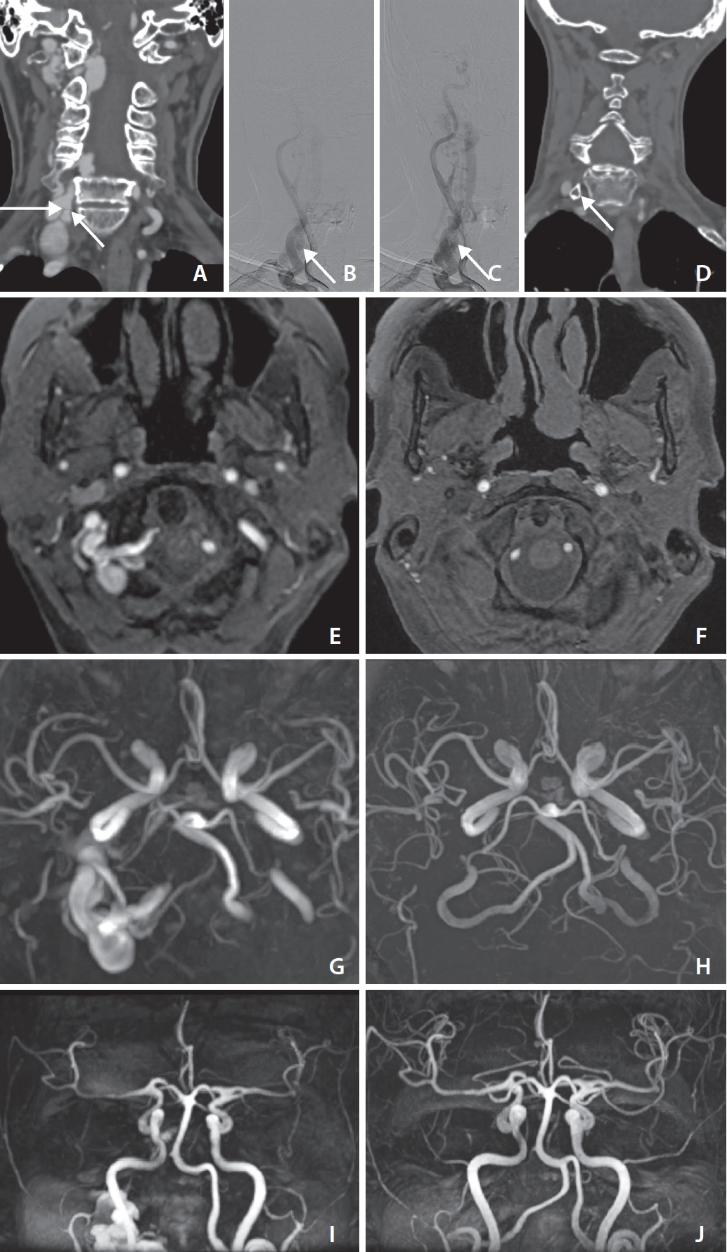
An iatrogenic vertebrovertebral fistula due to a puncture for central catheterization was observed in computed tomography (CT) and magnetic resonance (MR) angiography images of a patient in their sixties who was hospitalized in the intensive care unit because of mitral valve replacement 8 years ago. The patient was taking antiaggregant/anticoagulant medication. The vertebrovertebral fistula (diameter: 5 mm) was fed from the right vertebral artery at the C7–T1 level and drained predominantly into the right vertebral/subclavian veins and internal vertebral plexus on digital subtraction angiography (DSA) images. After a 6-french guiding catheter (Sofia; MicroVention) was placed in the right vertebral artery, the fistulous point was crossed with a Via-33 catheter and a 0.014-inch guidewire with a soft tip. The largest WEB device (W2-11-S, 11×9.6 mm) was placed just distal to the fistulous point (Fig. 1). The following control series after 6 minutes showed complete occlusion of the fistula, and the device was successfully detached without migration or movement. Supplementary coil embolization was not performed. First-year follow-up CT and MR angiograms revealed total occlusion of the fistula and normal posterior circulation without any complications.
Case 2
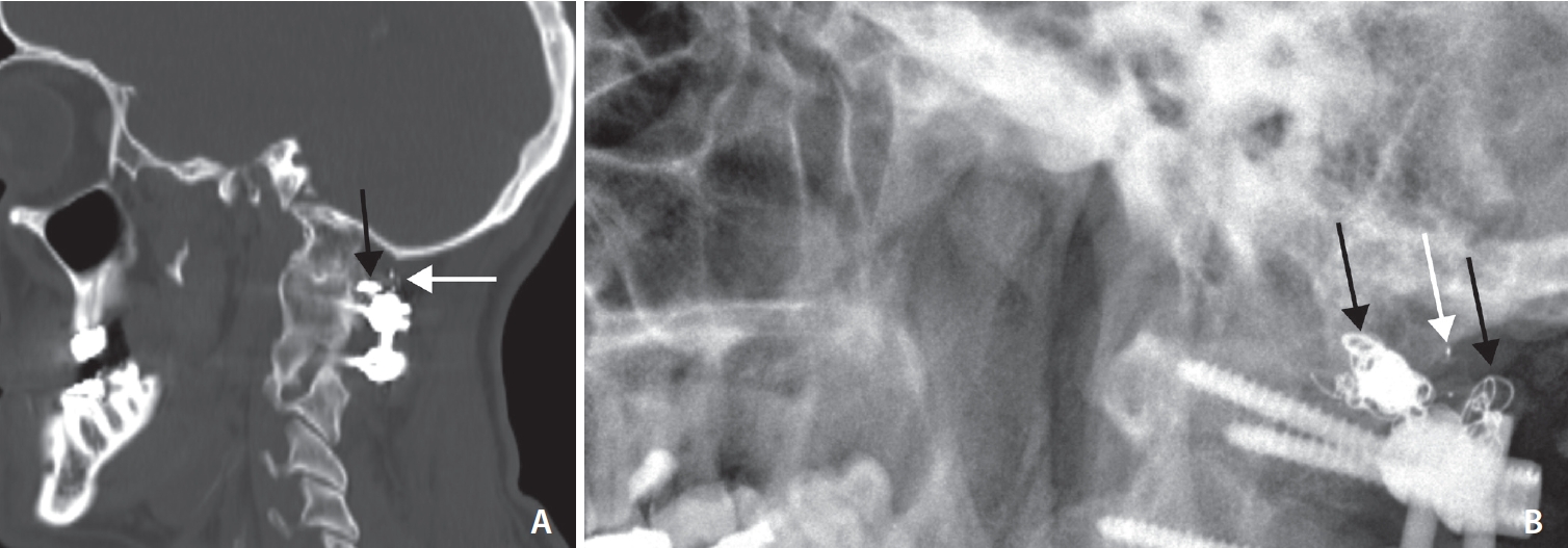
Our department was urgently contacted about a patient in their forties due to an intraoperative vertebral artery injury during spinal stabilization after a traffic accident. On DSA examination, an AVF originating from the left vertebral artery was observed at the C2 level. A WEB device with an 11 mm diameter (W2-11-9) was placed at the fistula level using the same approach as in case 1. Since the fistula was not completely closed on late-phase DSA, it was closed using a few coils as an adjunctive technique (Fig. 2). No recurrence was detected in the physical and radiological examinations of the patient performed 5 years later.
DISCUSSION
Endovascular management of head and neck AVFs is a more accurate option than surgery, with acceptable morbidity and mortality rates [2]. The WEB device has demonstrated good long-term occlusion rates and many advantages compared with other techniques for aneurysm embolization [3]. These advantages appear to be valid for embolization of AVFs or carotid-cavernous fistulas based on preliminary findings [4]. WEB-assisted embolization does not require antiplatelet loading unlike stenting, and WEB devices can be easily applied in emergency conditions [3,4]. Unlike with liquid embolic agents, there is no trans-collateral migration or microcatheter trapping risk for the WEB [5].
As applied in our cases, if the WEB device is larger than the diameter of the fistulous opening and the venous portion is just distal to the fistula, the AVF can close successfully without an additional technique or material. If the size of the available largest WEB device is equal to or smaller than the diameter of the fistula opening or venous portion immediately distal to the opening, supplementary coiling should be performed. In addition, in case the fistula cannot be closed on control runs 5–10 minutes after deployment of the WEB, coiling must be used as an adjunctive technique. Relapses may occur after treatment with WEB devices. No recurrence was observed in our 2 cases. CT angiography and 3-tesla non-contrast MR angiography are useful in the diagnosis and follow-up of these patients.